
Virus Scans
Virus scan
Human enteric viruses are considered one of the main causes of acute foodborne gastroenteritis. This is due to their high resistance to environmental conditions and to most treatments commonly used in the food industry. It is also due to their low infectious dose.
More recently, the SARS-CoV-2 (COVID-19) pandemic — an infectious virus — has highlighted the importance of controlling its presence and spread in our environment. Numerous scientific publications have highlighted the significant circulation of enteric viruses and SARS-CoV-2 in aquatic environments subject to discharges from wastewater treatment plants.
Our virus detection services
Rely on our molecular biology team
Virus detection and quantification
Main matrices can be analysed for viral contamination
Our virus detection services
Rely on our molecular biology team
Direct access to our biologists
Flexible tools and methods tailored to your requirements
Studies customised to meet your specific needs
Virus detection and quantification
Using quantitative PCR or quantitative RT-PCR techniques.
Highly specific, sensitive, and rapid methods, with results available within hours
Detection and quantification of a wide range of viruses, including GI and GII noroviruses, hepatitis A and E viruses, rotavirus A, adenovirus, enterovirus, astrovirus, sapovirus, aichivirus, cosavirus, salivirus, and SARS-CoV-2 (COVID-19)
Main matrices analysed for viral contamination
Drinking water (tap and bottled)
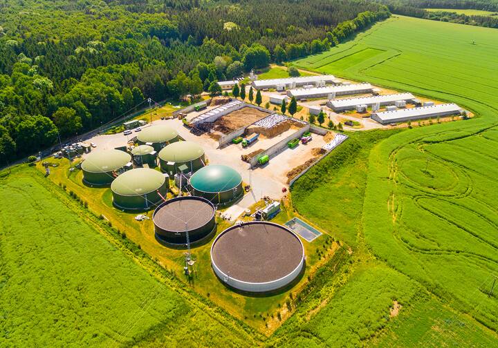
Wastewater treatment plant water (wastewater and effluent)
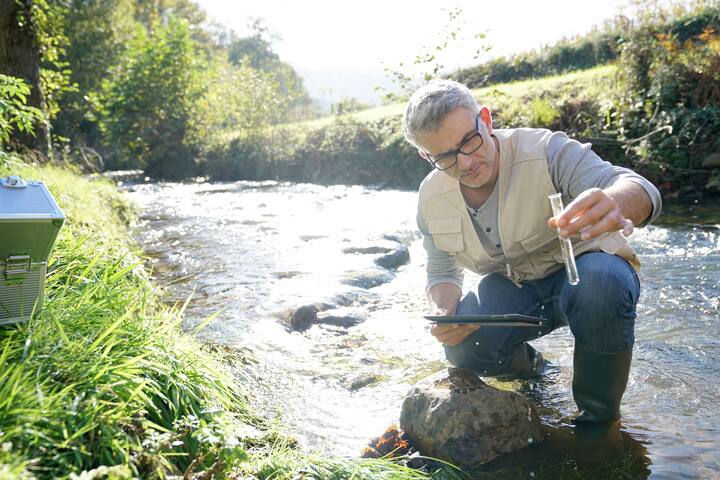
Natural bathing water (rivers and lakes)
Swimming pool water
Groundwater (wells and springs used to produce drinking water)
Sediments and sludge from wastewater treatment plants
Food matrices (shellfish, vegetables, fruit, meat)
Regulations
Regulation (EC) No 1441/2007 stipulates that “food shall not contain microorganisms or their toxins or metabolites in quantities that present an unacceptable risk to human health.”
Directive 2020/2184 of the European Parliament and of the Council of December 16, 2020, recasting the directive on the quality of water intended for human consumption, including somatic bacteriophages as indicators of faecal contamination in raw water.
Learn more about virus scanning
The risk of viral contamination mainly occurs at three levels:
that of commercialised foodstuffs and drinking water, whose safety is assessed upstream, within the framework of regulations;
that of the professional environment, which is also subject to specific regulations (Directive 2000/54/EC on biological agents at work);
and events to which individuals are exposed on a daily basis, whether accidental or not (direct exchange of biological fluids between humans, accidents involving contaminated environments or wild animals, etc.).
In addition to direct contact, the main sources of viral contamination are the airborne route and the food route when food or drinking water has been in direct or indirect contact with faeces. Controls, therefore, focus mainly on human enteric viruses (HEVs) and more specifically on three groups:
noroviruses, which cause most cases of gastroenteritis (diarrhoea, vomiting, abdominal pain)
hepatitis A and E viruses (inflammation of the liver)
rotaviruses, which are characteristic of gastroenteritis in children
Among infections, the food route is the most likely route of transmission due to the handling (mainly by humans) that food undergoes prior to distribution and the fact that, for the most part, it provides conditions that are conducive to maintaining the integrity of viruses (and therefore their infectiousness).
In addition, numerous scientific publications (Lee et al, 2014, López-Gálvez et al, 2016, Opere et al, 2020) highlight the significant circulation of enteric viruses in the environment, particularly in aquatic environments and, for the most part, downstream of wastewater treatment plant discharges.
However, the analysis of these results in terms of human health risk assessment must be qualified by the fact that:
in many studies, the detection of EVEs is that of their genome, which does not account for their infectiousness.
studies that address this aspect do so using surrogate organisms (phages, murine norovirus) and are therefore not easily transferable to human populations.
In summary, the frequency of gastroenteritis seems to be explained by both a certain resistance of VEH to environmental conditions and to many of the treatments commonly used in the food industry, but also by their low infectious dose.
A virus can be sought because of the danger it poses, but also as a direct or indirect indicator of faecal contamination. Somatic coliphages, for example, are sought as an indicator of the presence of a bacterium, Escherichia coli, which itself indicates that the water being analysed has come into contact with wastewater.
One of the classifications used for virus analysis distinguishes between three categories:
those that focus on their infectivity, which involve culture on a solid or liquid medium. Several methods are used to interpret the results: TCID50, immunofluorescence.
those that focus on their constituents, nucleic acids or proteins (qPCR, immunoblotting, immunoprecipitation, ELISA, hemagglutination)
direct counts in which viruses are considered as particles (flow cytometry, transmission electron microscopy)
Analyses also differ according to the phase of the protocol in which they are carried out:
extraction and concentration of viruses from the analysed medium (filtration, precipitation-centrifugation, etc.)
analysis of the extract itself (lysis ranges, molecular biology, microscopy, etc.)
Due to the time required for implementation and the cost of the necessary equipment, some of these techniques are more commonly used in research laboratories than in analysis laboratories.
1 – Nucleic acid-based techniques (known as molecular biology techniques)
This type of analysis involves three stages:
extraction and concentration of viruses from the medium being analysed
lysis of the viruses to extract nucleic acids (RNA or DNA, depending on the virus)
molecular analysis of the extracts: qPCR (DNA viruses) or RT-qPCR (RNA viruses)
Virus extraction phase. The ABIOLAB laboratory uses two techniques for extracting and concentrating viruses: filtration using an electropositive filter cartridge, and a technique combining elution, precipitation and centrifugation. The efficiency of extraction varies greatly depending on the composition of the water being analysed. This step remains the main weakness of the protocols, with extraction yields sometimes very low (<10%). It is the focus of some of the laboratory's R&D work. Nucleic acid detection and quantification phase (qPCR or RT-qPCR). qPCR is the abbreviation for “quantitative Polymerase Chain Reaction.” The aim of this technique is to multiply the DNA or RNA chains that have been extracted from the sample to be analysed (where they are, in principle, found in small quantities) so that their concentration exceeds the sensitivity threshold of the measuring device. The natural mechanisms of nucleic acid multiplication are reproduced in vitro in well plates in which the following compounds are brought into contact:
an unlimited quantity of the four nucleotides that make up nucleic acids
a heat-resistant DNA polymerase (Taq polymerase) to catalyse the synthesis
oligonucleotides called primers that flank the sequence to be amplified, the amplicon
in the technique known as Taqman, a sequence capable of pairing in this same region, onto which a fluorescent reporter and a fluorescence-suppressing compound (quencher) have been grafted. During each amplification cycle, the nucleotides that make up this probe are dissociated. This results in the separation of the quencher and the reporter. The latter then emits a fluorescent signal under the effect of UV radiation (Figure 1). The amplitude of this signal, therefore, increases proportionally with the number of cycles. The final value recorded, the Ct (cycle threshold), is the number of cycles required (and therefore the number of amplicons produced) for the signal to be above the background noise of the device at the start of the exponential phase. The number of cycles required to reach this threshold is therefore inversely proportional to the number of occurrences of the sequence in question in the initial sample. The amplification results appear in real time on the thermocycler screen, hence the name real-time qPCR given to this technique.
If the sequence to be amplified is RNA, it must first be transcribed into complementary DNA (cDNA) in order to benefit from the use of DNA polymerase (RNA polymerases do not have the same reaction properties). The starting mixture must therefore contain the corresponding enzyme, reverse transcriptase. The technique becomes RT-qPCR (Reverse Transcription Quantitative Polymerase Chain Reaction), in which the abbreviation RT could be repeated for its second meaning, the full name of the technique being “Reverse Transcription Real Time Quantitative Polymerase Chain Reaction.” The well plates are placed in a thermocycler where they are subjected to temperature variations whose amplitude and duration correspond to the optimal conditions for the successive stages of synthesis. The reactions occur spontaneously under the control of these temperature cycles. The cycle is repeated until the fluorescence signal reaches a plateau phase (Figure 1).Figure 1. Example of a graph produced by a thermocycler
The initial amount of amplicons in the analysed sample is calculated from calibration curves established beforehand, using known concentrations of the nucleotide sequence in question for which the Ct was measured under the same operating conditions.
2 – Techniques based on the infectious nature of viruses
These are used more specifically in laboratory research. Reading using the lysis range technique. In these techniques, cells that are sensitive to infection by the virus in question are cultured in flat flasks or Petri dishes until they are confluent. A known volume (the inoculum) of several dilutions of the sample to be analysed is deposited in several places on the cell layer. After an incubation period, the areas in which the virus concentration was sufficient to cause cell lysis appear as lighter circles (compared to the intact cell background). These areas are called lysis zones (Fig. 2). The initial concentration of the sample is calculated on the assumption that at least one viral particle is present in the highest dilution.
Detection and counting of viruses using the lysis zone technique
Flat culture flask
Petri dish
Lysis zones on bacterial mat
Reading by visual assessment of viral attack. The TCID50 (Tissue Culture Infectious Dose) is a form of expression of the titer of a viral suspension. It is the concentration of an inoculum at which 50% of the cells in an infected culture have been clearly affected (cytopathic effect or destruction) by the virus. The reading is visual (lighting of the culture). It can be automated if the culture has been done in wells, using a nephelometric device.
The Normec laboratory has made virology one of its areas of expertise, with the support of the ASPOSAN association. This focus, which is now widely recognised, has led to the signing of several collaboration agreements with two nearby universities (Université Grenoble Alpes and Université Savoie Mont Blanc) and with the Rhône-Alpes Auvergne Region. Two PhDs are currently being prepared in this context. The work of the R&D unit is focused on improving techniques for extracting and concentrating viruses from environmental water and on researching innovative techniques in this field. It also aims to measure and better understand the effect of the various stages of wastewater treatment in treatment plants on viral loads.
Bibliographic references
Zárate S, Taboada B, Yocupicio-Monroy M, Arias CF. Human Virome. Arch Med Res. 2017 Nov;48(8):701-716.
Opere WM, John M, Ombori O. Occurrence of Enteric Viruses in Surface Water and the Relationship with Changes in Season and Physical Water Quality Dynamics. Adv Virol. 2020.
López-Gálvez F, Truchado P, Sánchez G, Aznar R, Gil MI, Allende A. Occurrence of enteric viruses in reclaimed and surface irrigation water: relationship with microbiological and physicochemical indicators. J Appl Microbiol. 2016.
Chang Soo Lee, Cheonghoon Lee, Jason Marion, Qiuhong Wang, Linda Saif, Jiyoung Lee. Occurrence of human enteric viruses at freshwater beaches during swimming season and its link to water inflow, Science of The Total Environment,472, 2014: 757-766.
These services might also be of interest to you

Hydrobiology
Wastewater sampling and analysis
Leading companies in this service

Normec Abiolab
Montbonnot-Saint-Martin France
Curious to know what we can do for you?
FAQ
Frequently asked questions
A virus scan detects and quantifies human enteric viruses and other infectious viruses in water, food, and environmental samples. It helps identify contamination risks and ensures public health safety.
Virus scans are crucial for preventing infections from viruses like norovirus, hepatitis A and E, rotavirus, and SARS-CoV-2. They also monitor viral contamination in water and food, helping to control outbreaks and protect public health.
Viruses are detected using sensitive molecular biology techniques such as qPCR and RT-qPCR, or by assessing their infectivity through culture-based methods. These methods provide rapid, accurate, and reliable results.